Vanderbilt Health receives $3.4 million grant to advance breast cancer imaging

Submitted by vicc_news on

Vanderbilt Health has received a five-year, $3.4 million Academic-Industrial Partnership grant from the National Cancer Institute to support development of magnetic resonance (MR) cytometry as a new tool for breast cancer imaging.

MR cytometry is a magnetic resonance imaging technology pioneered by Vanderbilt Health investigators that provides information about tumor microstructure which cannot be obtained from conventional MRI.

Junzhong Xu, PhD, Director of Cancer Imaging Research at the Vanderbilt University Institute of Imaging Science (VUIIS), is principal investigator of the project. He will work with VUIIS Director John Gore, PhD, and with grant co-investigator Ryan Robison, PhD, MR Clinical Science Leader for North America at Philips Healthcare.

The investigators will integrate the technique into existing clinical MRI systems and develop a quality control system to support consistent use across scanners and institutions. They will then work with researchers from the University of Washington in Seattle to evaluate this technology in clinical breast cancer imaging.

This project “represents an important step in translating a Vanderbilt-developed imaging technology into a tool that could eventually be used in routine clinical care,” Xu said. “Working closely with Philips allows us to address the technical challenges required to move the method beyond a research environment and toward broader clinical use.”

Despite significant advances in early detection and treatment in recent years, breast cancer remains a leading cause of cancer mortality in women. Tissue biopsies provide essential diagnostic information but may provide an incomplete picture of the tumor and are difficult to repeat frequently over the course of treatment.

MR cytometry uses advanced diffusion MRI technology, including oscillating gradient spin echo, to probe how water molecules move within tumors at micrometer length scales. Advanced data acquisition and analysis of this movement enable researchers to estimate mean cell size and cell density noninvasively.

Combining biopsy findings with MR cytometry could give clinicians a more complete picture of a tumor and potentially help guide treatment decisions.

“The ability to measure cell size, cell density and other microstructural features using oscillating gradient diffusion imaging is immensely exciting, given its potential to monitor disease progression and inform clinical decision-making,” Robison said.

“Working alongside Dr. Xu and the team at VUIIS is a tremendous privilege, as they are the pioneers of this technique and are widely recognized as the world leaders in its development, validation and clinical translation,” he said.

“We have had a strong relationship with Philips for over 20 years, and it is encouraging to obtain funding that will be of mutual benefit and help patients,” said Gore, University Distinguished Professor of Radiology and Radiological Sciences, and holder of the Hertha Ramsey Cress Chair in Medicine.

Xu, Associate Professor of Radiology and Radiological Sciences, said MR cytometry has the potential to be applied to imaging other cancers and diseases in which changes in cellular microstructure are important.

The NCI Academic-Industrial Partnership grant (R01CA308592) supporting the research is designed to move promising technologies toward clinical use through collaborations between academic and industry partners.

The post Vanderbilt Health receives $3.4 million grant to advance breast cancer imaging appeared first on Vanderbilt Health News.
Vanderbilt Health performs nation’s first breast cancer surgery using intraoperative PET-CT scan technology

Submitted by vicc_news on

Following the introduction of a surgical protocol that leveraged intraoperative imaging with a combination PET-CT scanner to assess the success of head and neck cancer resection, surgeons have used the technology for breast cancer.

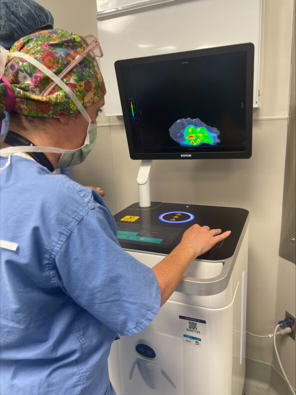
Denise Garcia, MD, uses the Xeos Aura 10 scanner to image a recently excised breast tumor from a patient. The use of the Xeos Aura 10 device ensures the imaging and assessment can be done in the operating room rather than in the pathology lab. (photo by Kyrionna Golliday)

Investigators led by Michael Topf, MD, Associate Professor of Otolaryngology-Head and Neck Surgery, performed the nation’s first surgery using intraoperative PET-CT scanning in September 2025. Now, a surgical team led by Denise Garcia, MD, Assistant Professor of Surgery in the Division of Surgical Oncology and Endocrine Surgery, has applied the technology to successfully resect a breast cancer mass.

“This application of intraoperative PET-CT is proof that countless patients can benefit from the expansion of this novel imaging methodology,” said Garcia. “Our team is proud to apply it to a type of tumor that has not yet been imaged for the purposes of assessing margin status. As our institution expands treatment methodologies to more types of cancer, we can cure more patients and give them peace of mind that their surgery has been completed with precision. This technological advancement underscores the success we’ve had across multiple disciplines in working toward that goal.”

Rapid expansion of intraoperative PET-CT scanning boosts efficiency, offers peace of mind to surgeons and patients alike

With each new application, surgeons are demonstrating that an intraoperative PET-CT imaging protocol can help reduce wait time for results from several days to a matter of minutes and allow surgical teams to know immediately whether they need to continue operating.

During surgery, the patient receives a dose of a radioactive agent that illuminates the cancer tissue in the scanner. Once the tumor is excised, it is placed in a specialized mobile PET-CT scanner called an Aura 10 device, developed and supplied by Belgium-based surgical technology company Xeos. The scanner negates the need to send the specimen to the pathology lab, providing surgical teams with a real-time view and allowing them to quickly determine if the entire cancerous mass was removed.

If any mass remains, the operation continues. If the cancer has been successfully resected, the surgery concludes, and the patient is sent home with peace of mind, knowing they won’t need to return for a follow-up surgery, and with confidence that their surgeons have a precise, immediate look at the results of the surgery. Because patients receive the radioactive agent on the day of surgery rather than in advance, they also receive a lower dose of radiation.

The post Vanderbilt Health performs nation’s first breast cancer surgery using intraoperative PET-CT scan technology appeared first on Vanderbilt Health News.

Liquid biopsy predicts response to breast cancer immunotherapy

Submitted by vicc_news on
Justin Balko, PharmD, PhD
Justin Balko, PhD, PharmD

Immunotherapy has become a standard of care in treating high-risk, early-stage breast cancers, yet it has had limited success in shrinking tumors. New biomarkers that can improve outcomes for patients are urgently needed.

Now, a study led by researchers at the Vanderbilt-Ingram Cancer Center has found that repeated blood sampling — essentially, a liquid biopsy — can assess and predict the evolving antitumor immune response to therapy.

This minimally invasive and cost-effective alternative to tissue biopsy offers “an accessible tool for tailoring treatment strategies in breast cancer,” they reported April 22 in the journal Science Translational Medicine.

The researchers performed RNA sequencing on 546 peripheral blood samples from 160 patients with high-risk, stages 2 or 3 breast cancers negative for human epidermal growth factor receptor 2 (HER2) during treatment with either chemotherapy alone or in combination with immunotherapy.

Justin Balko, PhD, PharmD, professor of Medicine and Pathology, Microbiology and Immunology at Vanderbilt Health and the paper’s corresponding author, acknowledged several co-authors — investigators from the nationwide I-SPY2 clinical trial — who, among other contributions to the study, provided the blood samples.

Co-author Laura Esserman, MD, MBA, director of the Breast Care Center at the University of California, San Francisco, is principal investigator of the I-SPY2 trial, which is assessing novel treatment strategies for subsets of breast cancer based on their molecular characteristics (biomarker signatures). Vanderbilt Health is among 42 trial locations.

Cell-free DNA testing, another form of liquid biopsy, is routinely used clinically for detection, diagnosis and therapeutic monitoring of a variety of malignancies.

Balko and his colleagues sampled the transcriptome, the transcription of genes involved in the clonal expansion and activation of antitumor immune cells called T cells. They found it predicted response to the immunotherapy drug pembrolizumab.

While validation is needed, this new liquid biopsy has the potential to “guide immunotherapy decision-making, tailor treatment regimens, and advance precision oncology, not only in (breast cancer) but potentially in other solid tumors as well,” the researchers concluded.

The paper’s first author, Xiaopeng Sun, PhD, is now at Merck. Co-authors at Vanderbilt Health are Andres Ocampo, Jacey Marshall and Julia Steele, graduate students in the Vanderbilt Program in Cancer Biology, and Susan Opalenik, PhD, senior research supervisor in the Balko lab.

The study was supported in part by National Institutes of Health grants P50CA098131, PO1CA210961, R01CA255442, U54CA274502, P30CA082103, P30CA068485 and NIH/NCI Imaging grant 28XS197 P-0518835), a Department of Defense Era of Hope Award, the Breast Cancer Research Foundation, Breast Cancer Research — Atwater Trust, Stand Up To Cancer, the California Breast Cancer Research Program and Give Breast Cancer the Boot.

The post Liquid biopsy predicts response to breast cancer immunotherapy appeared first on Vanderbilt Health News.

Genomic testing finds a higher prevalence of high-risk breast cancer in Black women

Submitted by vicc_news on
Sonya Reid, MD, MPH

Black women in the United States are less likely than white women to be diagnosed with breast cancer, yet they are more likely to die from the disease.

In a study of more than 1,000 women, researchers at Vanderbilt Health and Agendia, which specializes in genomic testing, found that Black women were more likely to have early, high-risk breast cancer that is hormone receptor positive (HR+) and negative for the human epidermal growth factor receptor 2 (HER2-).

These findings, reported March 19 in the Nature partner journal npj Breast Cancer, highlight the critical need for tumor genomic testing for all patients to identify those with high-risk tumors, which occur more frequently in Black women, and which require more aggressive treatment to prevent recurrence.

“By moving beyond traditional clinicopathologic features and incorporating genomic classification, we can more accurately identify biologically aggressive disease and tailor more precise, personalized treatment,” said the paper’s corresponding author, Sonya Reid, MD, MPH, associate professor of Medicine at Vanderbilt Health.

“The risk of recurrence of breast cancer in Black women has often been underestimated by traditional clinical features, driven largely by their underrepresentation in clinical trials,” study co-author William Audeh, MD, Agendia’s Chief Medical Officer, said in a news release.

“By providing a genomic assessment of tumor biology, we can ensure that women with breast cancer will receive individualized care that improves their long-term outcomes,” Audeh said. The collaboration with Vanderbilt Health “underscores our shared commitment to bringing precision medicine to all women.”

The research included 1,018 women with HR+, HER2- early-stage breast cancer who were enrolled in the Breast Cancer Etiology, Survival and Treatment Outcomes (BEST) study, and the Full-genome Data Linked with Clinical Data to Evaluate New Gene Expression Profiles (FLEX) study.

The BEST study is a population-based observational cohort of women identified through state cancer registries in Florida and Tennessee who were diagnosed with breast cancer between 2005 and 2015. FLEX is a multicenter, prospective observational study of genomic profiling and its impact on prognosis, treatment decisions and clinical outcomes.

The researchers applied two RNA-based technologies developed by Agendia to evaluate tumors: “MammaPrint,” which analyzes the 70 most important genes associated with breast cancer recurrence, and “BluePrint,” which classifies tumors into subtypes based on an expression profile of 80 different genes.

They found that Black women had a higher proportion of genomically high-risk luminal B and basal-type tumors compared to white women and that survival at three years was determined by the molecular subtype of their tumors.

“Understanding the biological and tumor genomic differences by race could improve treatment decisions and promote optimal care for Black females with early-stage breast cancer,” the researchers concluded, “ultimately improving long-term outcomes.”

Co-authors from Vanderbilt Health were Lindsay Venton, Jennifer Whisenant, PhD, Anne Weidner, MPH, and Tuya Pal, MD.

The post Genomic testing finds a higher prevalence of high-risk breast cancer in Black women appeared first on Vanderbilt Health News.

Join Nicole Kidman in supporting breast cancer research and care

Submitted by vicc_news on

Actress Nicole Kidman knows the impact when someone you love has cancer. For her, it was her mother, when Kidman was only in her teens.

Kidman’s mother lived another 40 years, for which she is grateful. The experience caring for her mother — and being able to have her mother for decades more — drives Kidman’s commitment to advancing breast cancer research.

Kidman recently donated her time, voice and influence to support breast cancer research at Vanderbilt-Ingram Cancer Center. She recorded a video alongside her friend Vandana Abramson, MD, co-leader of Vanderbilt Ingram’s Breast Cancer Research Program, sharing her story for Breast Cancer Awareness Month and beyond. The video has been posted on VICC Facebook and Instagram channels, and Kidman amplified it with her 25 million fans on Instagram and Facebook.

“We’re so grateful for the support of Nicole Kidman in raising awareness for breast cancer research at Vanderbilt-Ingram Cancer Center,” Abramson said. “Nicole witnessed her mother’s journey as a breast cancer patient, and this has inspired her to help others find healing and strength. You can stand with Nicole — and stand with us — in the fight against breast cancer. Your gift fuels research that leads to breakthroughs. It helps us personalize treatments, reduce side effects and offer hope where there was once uncertainty. It brings us closer to a world where breast cancer is no longer a life-altering diagnosis.”

We hope you’ll watch the video and share it with your family and friends on your own social media, email or other channels. To see the video, learn more and help make an impact, visit GiveToVICC.org.

In addition to this video, Kidman has also donated her time and support to be “the voice” for Vanderbilt Health’s upcoming brand advertising campaign, which will launch later this fall.

The post Join Nicole Kidman in supporting breast cancer research and care appeared first on VUMC News.

Subscribe to Breast_Cancer