Following the introduction of a surgical protocol that leveraged intraoperative imaging with a combination PET-CT scanner to assess the success of head and neck cancer resection, surgeons have used the technology for breast cancer.
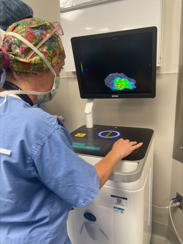
Denise Garcia, MD, uses the Xeos Aura 10 scanner to image a recently excised breast tumor from a patient. The use of the Xeos Aura 10 device ensures the imaging and assessment can be done in the operating room rather than in the pathology lab. (photo by Kyrionna Golliday)
Investigators led by Michael Topf, MD, Associate Professor of Otolaryngology-Head and Neck Surgery, performed the nation’s first surgery using intraoperative PET-CT scanning in September 2025. Now, a surgical team led by Denise Garcia, MD, Assistant Professor of Surgery in the Division of Surgical Oncology and Endocrine Surgery, has applied the technology to successfully resect a breast cancer mass.
“This application of intraoperative PET-CT is proof that countless patients can benefit from the expansion of this novel imaging methodology,” said Garcia. “Our team is proud to apply it to a type of tumor that has not yet been imaged for the purposes of assessing margin status. As our institution expands treatment methodologies to more types of cancer, we can cure more patients and give them peace of mind that their surgery has been completed with precision. This technological advancement underscores the success we’ve had across multiple disciplines in working toward that goal.”
Rapid expansion of intraoperative PET-CT scanning boosts efficiency, offers peace of mind to surgeons and patients alike
With each new application, surgeons are demonstrating that an intraoperative PET-CT imaging protocol can help reduce wait time for results from several days to a matter of minutes and allow surgical teams to know immediately whether they need to continue operating.
During surgery, the patient receives a dose of a radioactive agent that illuminates the cancer tissue in the scanner. Once the tumor is excised, it is placed in a specialized mobile PET-CT scanner called an Aura 10 device, developed and supplied by Belgium-based surgical technology company Xeos. The scanner negates the need to send the specimen to the pathology lab, providing surgical teams with a real-time view and allowing them to quickly determine if the entire cancerous mass was removed.
If any mass remains, the operation continues. If the cancer has been successfully resected, the surgery concludes, and the patient is sent home with peace of mind, knowing they won’t need to return for a follow-up surgery, and with confidence that their surgeons have a precise, immediate look at the results of the surgery. Because patients receive the radioactive agent on the day of surgery rather than in advance, they also receive a lower dose of radiation.
Gayle Knoop doesn’t post about her cancer on social media, so most people didn’t even know she had been diagnosed with Stage 4 colon cancer with liver metastases in October 2024, or that the cancer had returned after nine months, or that she was preparing for Christmas 2025 to be her last with her family.
She was trying to stay upbeat.
“I’m at home over the holidays thinking this is probably my last Christmas with my family,” said Knoop, who has a husband, 22-year-old daughter, three dogs, and an older brother. “It was miserable.”
After treatment, she had been cancer-free for nine months, but the cancer returned in her liver. Her hometown doctor told her the clinical team had met about her case, and she wasn’t a candidate for surgery. She could do Y90 radiation, which she did not want to do.
Knoop said she finally “dug deep” and started going down all the rabbit holes and doing her research.
It was different than the pressures she felt growing up playing volleyball, softball and basketball. This one was medical, and she felt hopeless, but she had a hunch there could be something out there for her.
“I want to be here for my daughter. I want to see her graduate from medical school. I want to see her get married,” she said. “And I’m like, OK, get up off your ***, start digging. You’ve done it before. You can do it again. And it led me to Vanderbilt. And, to me, that was my answered prayer for God to put the right people in my path, and he did.”
A better option than expected
With a little sleuthing and perseverance, she found something online with good reviews about Sekhar Padmanabhan, MD, just two-and-a-half hours away from her home in Louisville, Kentucky, at Vanderbilt-Ingram Cancer Center in Nashville. And it was in network for her insurance.
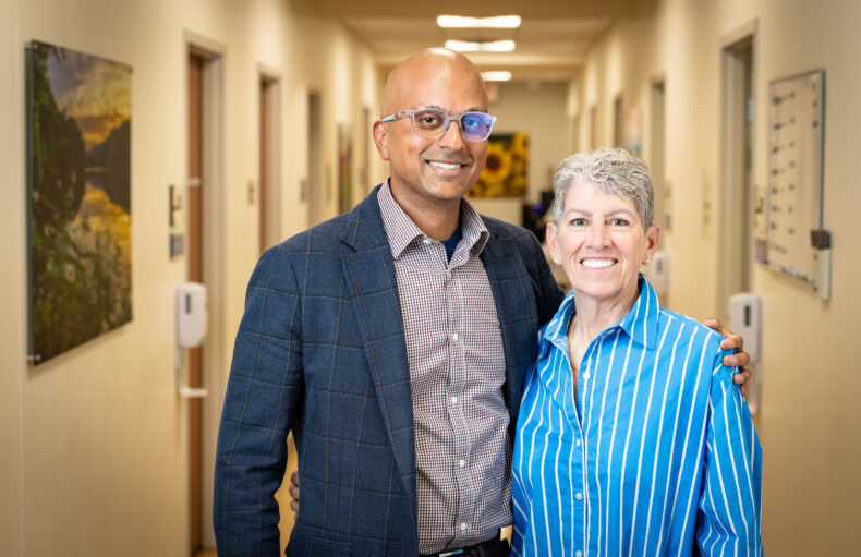
She came to Vanderbilt-Ingram in search of the histotripsy surgery she read about online, but Padmanabhan instead performed robotic-assisted laparoscopic surgery on Knoop to remove the left lobe of her liver and two tumors from the right lobe.
“The patient sheet just said she was there to discuss histotripsy, which she had read about online. Histotripsy was not what she needed; we had a better option than that,” said Padmanabhan, Assistant Professor of Surgery, who came to Vanderbilt-Ingram in 2021 to help build the robotic liver and pancreas program.
“Obviously she was shocked when I said, ‘I think we can remove these tumors and potentially put you on a path for a curative-intent option,’” he said.
Prior to 2022, there had not been a robotic liver resection done at Vanderbilt Health or in Nashville. There have now been more than 100 robotic liver resections at Vanderbilt Health.
A word no one says with Stage 4 cancer
Sekhar Padmanabhan, MD, and his patient Gayle Knoop, who traveled from Louisville, Ky., for treatment and found hope. (photo by Erin O. Smith)
Padmanabhan said Knoop’s story amplifies the importance of educating not only community oncologists but also patients about new therapies and technologies available to them.
“Gayle has a certain level of grit and is someone who will not be a defeatist … would not take ‘no’ for an answer. I am thankful that mentality is a part of her, because otherwise she wouldn’t have ended up here,” he said.
“As I was looking through her labs and her scans and her history, in my mind, I was saying, ‘What are we missing here? Why are we not operating on her? She has a chance at a curative option, and why are we not offering that?’”
Knoop required a couple of visits to Vanderbilt-Ingram to repeat scans, make sure her blood work looked good, and make sure her liver was healthy before she could have the robotic-assisted liver resection.
“He offered me a 20% chance of a cure. You know, nobody ever says ‘cure’ with Stage 4 cancer, and he offered me a 20% chance at a cure,” she said. “And, for the first time, I had hope, and it wasn’t false hope. I had hope, and I had confidence in the people that I was dealing with.”
The surgery also allowed her to go back to Louisville the next day instead of a more invasive surgery that would have included five days in an intensive care unit and a roughly 10-day hospital stay.
“These successes need to be shared with the world”
Padmanabhan said he saw her in the clinic two to three weeks after her surgery, and she looked great and was back to her usual day-to-day activities. At a later follow-up with Justin Lo, MD, PhD, Assistant Professor of Medicine, her scans and blood work continued to look great.
“When an institution and the people who make it up are this special, it deserves to be recognized,” Knoop said. “Tunnel vision is inevitable when this is your day-to-day life, but I truly believe Dr. Padmanabhan and his team at Vanderbilt-Ingram saved my life and could save the life of so many others. It is imperative that these successes are shared with the world.
“I was more than a patient or cancer case. I was a wife, mom and sister who needed his incredible knowledge of advanced technology and his surgical skills to spend more time in these roles with my family,” she said.
Padmanabhan said Vanderbilt-Ingram is “pushing the envelope” with new technologies like histotripsy and new programs like robotic liver surgery and robotic pancreas surgery, serving patients not only from Tennessee, but also parts of Kentucky, Alabama and Georgia.
“Gayle’s story is obviously unique to her and very personal for her but, unfortunately, that is the story that we hear a lot, especially in this area,” he said.
“Folks don’t necessarily have access to that care close to their home, so I would encourage people to see if there are newer drugs, treatments, surgeries, therapies and technologies that we can offer you that could extend your life, perhaps cure you in certain instances, but also ensure that we do all those things while maintaining your quality of life.”
Vanderbilt Health and Bertis, an artificial intelligence-driven proteomics-based precision medicine company, have announced a joint research and co-development collaboration. The endeavor marks a significant milestone in oncology by advancing the convergence of AI, spatial biology and translational cancer research.
By integrating Vanderbilt Health’s Molecular AI Initiative capabilities with Bertis’ proprietary deep proteomics and AI-enabled target discovery technologies, the collaboration will build an advanced, spatially resolved dataset to identify novel therapeutic targets and predictive biomarkers.
Traditional target discovery often relies on bulk tissue analysis, which loses the critical context of how cells are organized within a tumor. Vanderbilt Health’s Molecular AI approach changes this paradigm by employing sophisticated computational spatial analysis to generate high-resolution spatial molecular maps. This AI-driven spatial biology allows researchers to visualize and decode the complex architecture of the tumor microenvironment, specifically identifying how tumor, immune and stromal (connective tissue) cells interact in biologically and therapeutically relevant regions. By mapping the precise locations and spatial relationships of these cells, the Molecular AI platform can isolate the key cell populations responsible for treatment response or resistance.
These advanced spatial insights are then integrated with Bertis’ cutting-edge proteomics capabilities. While Vanderbilt Health maps the critical spatial context, Bertis will conduct deep proteomic and metabolomic profiling, applying its proprietary AI-enabled computational models to prioritize the most viable, druggable targets.
Tae Hyun Hwang, PhD
The initial focus of this joint research will be on HER2-low tumors (cancers that express low levels of the growth-promoting protein HER2), a historically challenging clinical area, with the potential to expand into additional tumor types based on data outcomes and joint scientific discussions. By layering spatial context over proteome-level data, the teams aim to pinpoint cell surface proteins that are uniquely positioned for emerging therapeutic modalities, including antibody-drug conjugates and cell-based therapies.
This sophisticated AI-driven spatial multimodal and deep proteomics pipeline is spearheaded by Tae Hyun Hwang, PhD, professor of Surgery, founding director of Molecular AI Initiative and director of AI Research in the Section of Surgical Sciences at Vanderbilt Health. Hwang also co-leads gastric cancer atlas efforts within the National Cancer Institute-funded Human Tumor Atlas Network (HTAN) and is spearheading international HTAN collaborations with South Korea’s National Cancer Center.
Highlighting the clinical necessity of this integrated approach, Hwang said, “Identifying therapeutic targets and understanding treatment response require a precise view of proteins, spatial context and tumor biology. By combining Vanderbilt Health’s Molecular AI and spatial analysis capabilities with Bertis’ proteomics and AI-enabled target discovery platform, this collaboration is designed to generate high-confidence therapeutic targets and predictive biomarkers that can support future translational research and therapeutic development.”
Bertis is led by co-CEOs Dong-young Noh and Seung-man Han, who emphasized the collaboration accelerates the global reach of their platform.
“Collaborating with Vanderbilt Health, a leading U.S. academic medical center with strong expertise in Molecular AI, spatial biology and cancer research, is highly meaningful and reflects the growing global recognition of Bertis’ technological capabilities,” Han said. “Through this collaboration, we aim to expand the role of AI-driven proteomics in drug discovery and identify therapeutic targets that may open new possibilities in oncology.”
Vanderbilt Health recently performed its 100th histotripsy, a noninvasive procedure in which highly focused ultrasound waves are directed at liver tumors to destroy cancer without ever making an incision.
The recipient, Aaron Davis of Cleveland, Tennessee, had just celebrated his 52nd birthday days before the procedure and was surrounded in the Vanderbilt University Hospital operating room by a surgical team he’s come to greatly admire.
Sekhar Padmanabhan, MD, assistant professor of Surgery, performed the procedure, in which a tub of water held over Davis’ abdomen served as the medium through which the ultrasound waves passed. In histotripsy, the focused ultrasound energy causes small gas bubbles in the tissue to rapidly expand and contract. This process forms a “bubble cloud,” forcing the targeted tumors to be liquified while avoiding damage to other tissue.
“Histotripsy is a novel procedure, but one that shows a great deal of promise,” said Padmanabhan. “Thanks to generous philanthropic support, we’re building a world-class program to continue offering this technology to patients who can benefit from a noninvasive surgical option that yields excellent results.”
Vanderbilt-Ingram Cancer Center is among the first institutions to offer histotripsy. Appealing to patients for its noninvasive nature, it avoids many of the traditional drawbacks of surgeries that use incisions, including pain management.
“When I had my liver resection, that was some of the worst pain I’ve ever felt in my life,” said Davis. “And I’m allergic to many pain medications, too. Being able to get put to sleep for surgery and wake up without the pain of having my abdomen cut open changes everything.”
Davis had previously been in Padmanabhan’s care to receive a hepatic artery infusion pump, which successfully delivered high doses of chemotherapy to his liver while minimizing toxicity to the rest of his body. And although the cancer returned, Davis knew he was in good hands with Padmanabhan and Kristen Ciombor, MD, MSCI, associate professor of Medicine in the Division of Hematology and Oncology, who eventually helped him settle on histotripsy as the best option to treat his latest recurrence of cancer.
“The doctors at Vanderbilt-Ingram Cancer Center changed my life,” said Davis. “Having a care team who knew exactly what I needed and recognized that a newer procedure could help me has given me hope that I can continue fighting cancer.”
Anna Means, PhD, whose 25 years of research at Vanderbilt Health advanced the understanding of early pancreatic cancer, died Nov. 28 at her sister’s home in Pelham, Alabama, following a 2023 diagnosis of brain cancer. She was 63.
A longtime collaborator of the late R. Daniel Beauchamp, MD, former chair of the Section of Surgical Sciences, in 2024 Dr. Means moved to the Department of Plastic Surgery where, as research professor of Plastic Surgery and Cell & Developmental Biology, she helped oversee development of a tissue engineering laboratory.
“Dr. Anna Means was my close friend, collaborator and colleague for over 30 years,” said Maureen Gannon, PhD, professor of Medicine in the Division of Diabetes, Endocrinology and Metabolism.
In addition to her research, Dr. Means mentored dozens of undergraduate, graduate and postdoctoral students, research staff and faculty. She was also a highly knowledgeable bird watcher, talented gardener and outdoor enthusiast. “She inspired us all with her grace and positivity and love of life,” Gannon said. “I will miss her terribly.”
An outstanding independent scientist and valued colleague, Dr. Means “was exacting and thorough in her scientific efforts and had an exceptional sense of integrity,” said Seth Karp, MD, H. William Scott Jr. Professor of Surgery and chair of the Section of Surgical Sciences. “She was highly respected across our campus and in the scientific community for her honesty, compassion and intelligence.”
A native of Ohio, Dr. Means earned her doctorate in Cell and Molecular Biology from the University of Wisconsin-Madison in 1991 and did postdoctoral work at Cornell University Medical College and Vanderbilt University before joining the Vanderbilt faculty in the Department of Surgery in 2000.
For 10 years until Dr. Beauchamp’s death in 2022, she was a close collaborator, serving as senior scientist in his lab, overseeing the work of research staff, and contributing as co-investigator and co-author to research that yielded important insights into the development of colorectal cancer.
In collaboration with other Vanderbilt faculty including Gannon and Christopher Wright, DPhil, professor of Cell & Developmental Biology, Dr. Means also led a highly productive research effort in pancreatic cancer and development of the pancreas.
She was founder and organizer of the Vanderbilt Pancreatic Cancer Researchers group, which convened a monthly research conference for basic and clinical investigators studying pancreatic cancer, and she organized the Beta Cell Interest Group, which held weekly seminars on studies related to pancreas development and function.
In 2009 Dr. Means received a Vanderbilt-Ingram Cancer Center Impact Award for her contributions to cancer research.
“At her core, Dr. Means was kindhearted, compassionate and deeply committed academically,” Karp said. “She demonstrated tireless dedication and achieved significant contributions to oncologic research as well as to the critical research and education missions of Vanderbilt. She made everyone around her better for having known, admired and worked with her.”
Dr. Means is survived by her mother, Joan Means, brothers, Christopher (Kim), Peter (Liz), and Patrick (Pam), and sisters, Michele Dragga (Chuck) and Kirsten Means.
Mark Kelley, MD, MMHC, medical director of the Williamson County General Surgery Division in the Department of Surgery at Vanderbilt University Medical Center, has retired, effective Oct. 1, after 28 years of exceptional service and leadership.
“Dr. Kelley transformed the Division of Surgical Oncology and Endocrine Surgery into one of the nation’s largest and most productive academic surgical oncology programs,” said Carmen Solórzano, MD, John L. Sawyers Chair in Surgical Sciences and chair of the Department of Surgery. “His remarkable contributions span clinical excellence, innovative research and impactful education.”
Kelley, an associate professor of Surgery in the Division of Surgical Oncology and Endocrine Surgery, is lauded by his colleagues and surgical leaders for his contributions both at VUMC and nationally.
“Dr. Kelley’s career reflects an unwavering commitment to excellence in clinical care, research, education and leadership,” said Seth Karp, MD, H. William Scott Jr. Chair in Surgery and chair of the Section of Surgical Sciences. “His legacy has profoundly shaped surgical oncology at Vanderbilt and beyond. We are grateful for his dedication and leadership.”
In 1997, Kelley joined the VUMC faculty as an assistant professor in the newly established Division of Surgical Oncology and Endocrine Surgery and served as clinical director of the Vanderbilt Breast Center until 2005. His leadership skills led to his appointment as chief of the Division of Surgical Oncology and Endocrine Surgery in 2002, a role he held until 2015.
“As clinical director of the Vanderbilt Breast Center, he led its development and transition from a small practice in the Village at Vanderbilt to a comprehensive breast center at One Hundred Oaks,” said Solórzano. “In 2005, he passed the clinical directorship to Dr. Ingrid Meszoely, whom he recruited back to Vanderbilt after her surgical oncology fellowship. Today, the Vanderbilt Breast Center is one of the largest and most comprehensive programs in the United States.”
Recognizing the importance of focused training in breast surgical oncology, Kelley developed the framework for a breast surgical oncology fellowship. Expansion of the curriculum under the leadership of Mary Hooks, MD, MBA, and Ingrid Meszoely, MD, led to accreditation of the program by the Society of Surgical Oncology in 2016.
Under Kelley’s leadership, the Division of Surgical Oncology and Endocrine Surgery expanded from four surgeons to a multidisciplinary team of more than 20 surgeons, advanced practice providers (APPs) and research scientists. He also played a pivotal role in recruiting and mentoring key faculty who now serve as VUMC surgical and Vanderbilt-Ingram Cancer Center leaders, including Rondi Kauffmann, MD, MPH; Christina Bailey, MD, MSCI; Kamran Idrees, MD, MSCI, MMHC; Meszoely, Solórzano and others.
Kelley was a sought-after mentor throughout his career, and assisted numerous medical students, surgical trainees and junior faculty as they participated in projects and developed their own research. Under his leadership, the Division of Surgical Oncology and Endocrine Surgery was highly ranked, year after year, as a favorite learning environment for general surgery trainees. More than 30 residents completed surgical oncology fellowships during Kelley’s tenure, and many are leaders in the field today, including five current VUMC faculty members.
He was an early advocate for the integration of APPs into clinical roles. In 1999, he established training and mentorship programs for APPs specializing in breast health and surgical oncology, and these programs served as models for integrating APPs into surgical practices throughout VUMC. Today, there are 10 APPs practicing in inpatient and outpatient roles in the Division of Surgical Oncology and Endocrine Surgery.
Kelley was a surgical innovator and the first surgeon in Tennessee to perform sentinel lymph node biopsy for breast cancer and melanoma in 1997. This procedure has transformed the care of these cancers. Kelley developed an Institutional Review Board (IRB)-approved protocol to train surgeons on this technique, leading to the rapid and safe application of the new surgical procedure at VUMC and in the community.
Kelly was also integrally involved in the development of multidisciplinary clinical and research programs at Vanderbilt-Ingram Cancer Center. From 2000-2012, he served as chair of the VUMC Cancer Committee. This group monitors and reports cancer volumes and outcomes, guides quality improvement, and ensures compliance with national cancer treatment standards. During his tenure as chair, VICC was continuously accredited by the American College of Surgeons Commission on Cancer. The program was also routinely recognized as one of the top National Cancer Institute-accredited comprehensive cancer centers nationwide during that time.
Early in his career, Kelley had an independent laboratory that focused on translational research in melanoma tumor biology and contributed to the early development of immunotherapy for melanoma. He established the melanoma and cutaneous malignancy tissue repository in 2003. This IRB-approved research repository has collected tumor tissue samples from patients undergoing surgical resection or biopsy with paired clinical data from more than two decades. This invaluable resource has supported high impact basic and translational studies that have led to novel combinations of immunotherapy and targeted therapy being investigated in clinical trials today.
“Dr. Kelley is highly committed to the advancement of cancer care through research, and we are grateful that he will remain active in clinical and translational research with VUMC and VICC as professor of Surgery, retired, to continue to improve care for patients with cancer,” said Karp.
Cancer patients who scored lower on health literacy screening experienced higher all-cause mortality, according to a study published in the journal Cancer.
The study followed Vanderbilt-Ingram Cancer Center patients for a median of 3.1 years who had taken the Brief Health Literacy Screen. Patients who had high health literacy on the screening lived 9.4 months longer compared to those with low health literacy (score of nine or lower). The 9,603 patients in the retrospective cohort study were diagnosed with either prostate, lung, breast, renal, colorectal, brain, head and neck, bladder, pancreatic, liver, sarcoma or gastric cancer.
“Cancer care is extremely complex, and we highlight that health literacy is an important risk factor in terms of survival in one of the largest studies conducted evaluating the impact of health literacy and cancer survival,” said the study’s senior author, Kamran Idrees, MD, MSCI, MMHC, Ingram Professor of Cancer Research, professor of Surgery and chief of the Division of Surgical Oncology and Endocrine Surgery.
He further stated, “Since health literacy is a modifiable risk factor, it provides us an opportunity for real-time identification of patients with low health literacy to personalize care, provide health literacy sensitive resources, tailored instruction and education to improve their cancer care.”
The screening consists of three multiple-choice questions about patients’ comfort levels with understanding medical information and filling out hospital forms. A point system, ranging from one to five, is assessed according to answers to the questions.
Although the study did not seek to discover causal findings, such as direct links between patient mortality and patients’ ability to make informed decisions about treatment scenarios, the investigators surmised the difference in outcomes was likely multifactorial.
The investigators stated they endorsed the routine collection of health literacy information for patients diagnosed with cancer and that they encouraged the adoption of strategies to improve organizational health literacy in facilities that provide cancer care. They noted that not all cancer patients with low health literacy experienced worse outcomes. Observational studies for specific cancer types that assess health literacy are needed to evaluate interventions aimed at improving outcomes, they said.
Other Vanderbilt authors on the study included Kelvin Moses, MD, PhD, Julia Whitman, MS, and Sunil Kripalani, MD, MSc.
The investigators state that to their knowledge the study is the first to assess the association between health literacy and all-cause mortality among different cancer types.
The research received support from a Society of Surgical Oncology Foundation Investigator Award for a grant titled “Health Literacy and Cancer Outcomes.”
An early-stage clinical trial, supported by the Department of Defense, has demonstrated that the targeted cancer drug trametinib shows potential as an interventional therapy to reprogram precancerous gastric lesions, potentially preventing them from becoming malignant, and that it can be administered safely.
The results of the Phase 1 trial involving 15 patients, which were published recently in Gastroenterology, were pleasantly surprising, said James Goldenring, MD, PhD, professor of Surgery and of Cell and Developmental Biology at Vanderbilt University Medical Center.
The primary goal of this trial was to evaluate whether a low-dose, limited duration treatment of two weeks with trametinib would be safe for patients at risk for developing a second cancer after having undergone resection of a Stage 1 gastric cancer. The drug also showed promise that it could be the first therapeutic intervention against precancerous lesions in the stomach.
Endoscopies revealed that trametinib reversed metaplasia, which is an abnormal change of cells into ones that are non-native to the tissue and can progress to dysplasia, an irreversible change in cell development that can lead to cancer. While the 15 patients in the study had no evidence of recurrent cancer, they did have extensive metaplasia when they entered the study.
“I was pleasantly surprised at how much benefit we could see in the endoscopies after one month and one year; it really was pretty remarkable,” said Goldenring, the Paul W. Sanger Professor of Experimental Surgery.
The reversal of the metaplasia could be viewed in endoscopic images and was confirmed with biopsies.
“I think that’s almost more compelling than anything else in this study,” Goldenring said. “I honestly did not expect endoscopies to be that different, but they were.”
However, he noted that follow-up clinical trials with more participants are needed to further validate the drug’s efficacy. The only significant side effect among the participants was one patient with a mild increase in blood pressure after trametinib treatment that returned to normal after the patient stopped taking the drug.
The patients in the study were recruited from Japan, where the clinical trial was led by Sachiyo Nomura, MD, PhD, in collaboration with Goldenring. Trametinib is an inhibitor of the MEK signaling pathway. MEK, an abbreviation for the mitogen-activated extracellular signal-regulated kinase pathway, plays an integral role in the development of stomach cancer.
The study was supported by a $2.5 million Department of Defense Translational Team Science Award, which is also supporting another clinical trial in the United States with similar aims. The U.S. clinical trial will evaluate the effectiveness of pyrvinium, an existing medicine that has been used for the past 70 years to treat pinworms in children, for a new purpose — reversing metaplasia of stomach cells and killing dysplastic precancerous cells. Pyrvinium also blocks the MEK pathway.
While stomach cancer is one of the three leading causes of cancer-related deaths worldwide, its incidence is lower is the U.S. Nevertheless, it does occur more frequently among minority ethnic groups, and incidence has been rising among young women. DOD support for clinical trials reflects the increased incidence of stomach cancer in minority groups, which make up a higher percentage of the U.S. armed services than of the general population. In the U.S., most stomach cancers are diagnosed at late stages when they are more difficult to treat.
Goldenring said he hopes the MEK inhibitor study will spur more research into therapeutic interventions for people with precancerous lesions who are at high risk for cancer.
“I’m hoping that this is a direction that multiple researchers might take in the future to really change the dynamics of how we’re going to intervene so that people don’t develop cancer,” he said. “That’s a different mindset than we’ve had previously.”
Eunyoung Choi, PhD, associate professor of Surgery and of Cell and Developmental Biology, is a co-principal investigator of the pyrvinium study along with Katherine Garman, MD, associate professor of Medicine at Duke University. Choi is also a co-author of the study published in Gastroenterology.
Goldenring is supported by grants from the Department of Defense, a Department of Veterans Affairs Merit Review Award, and the National Institutes of Health (R01DK101332 and R01CA272687. Choi is supported by grants from the National Institutes of Health (R37CA244970 and R01CA272687), the Department of Defense, the American Association for Cancer Research, and an American Gastroenterological Association Robert & Sally Funderburg Research Award.
Aimal Khan, MD, assistant professor of Surgery at Vanderbilt University Medical Center, noticed the puzzled or anxious expressions of patients trying to fully comprehend what he was saying during preoperative consultations, so he devised visual aids — three-dimensional models of the lower digestive tract.
The 3D models allowed patients to easily distinguish the ascending colon from the sigmoid colon, along with other parts of the digestive system. Patients could actually see where the surgery would occur, and Khan noticed that they asked more questions, felt more confident and seemed less anxious. He devised a study to determine whether his personal observations were scientifically valid.
The study, which was published June 3 in JAMA Network Open, determined that the 3D models made patients feel they played a bigger role in decision-making and that their anxiety levels decreased.
The patients were scheduled for partial or complete colon and/or rectal resections for colorectal cancer, diverticulitis or inflammatory disease. Fifty-one patients participated in the study with 28 receiving consultations using the 3D models and 23 receiving conventional consultations. The patients in the 3D arm of the study reported a significantly higher involvement in shared decision-making and significantly reduced anxiety levels compared to the other patients.
Khan and five other Vanderbilt surgeons conducted the study from March 2022 to June 2023.
“Using 3D models during consultations allowed our patients to truly visualize their surgery, which not only empowered them to take an active role in decision-making but also significantly eased their anxiety. This approach has the potential to transform how we communicate complex information to our patients. We are currently working with surgeons from other specialties, including thoracic surgery, ENT and surgical oncology, to validate these findings in a multicenter randomized trial,” Khan said.
The findings are important because other studies have shown that improvements in shared decision-making are associated with reduced hospital stays, lower health care utilization, improvement in patient-reported health outcomes and fewer emergency department visits.
The 3D models used in the study were developed in collaboration with the Department of Radiology. The modular designs, which were made with 3D printing, allowed each segment of the colon and rectum to be magnetically detached and reattached.
To the knowledge of the study’s authors, this is the first randomized clinical trial to compare the effectiveness of a 3D-printed model with usual care on colorectal surgery patients’ involvement in decision-making, anxiety and education.
Other Vanderbilt researchers who authored the study are Danish Ali, MD, Shannon McChesney, MD, Michael Hopkins, MD, Molly Ford, MD, Roberta Muldoon, MD, Timothy Geiger, MD, MMHC, Alexander Hawkins, MD, MPH, Georgina Sellyn, MA, Hillary Samaras, RN, and Dann Martin, MD, MS.
Researchers at Vanderbilt University Medical Center using artificial intelligence have helped develop two technologies for improving cancer care.
One technology called MSI-SEER, described in a study published in npj Digital Medicine, better predicts microsatellite instability-high status from standard pathology slides and provides clinicians with specific data, including any uncertainties with predictions. The other technology, a breakthrough three-dimensional imaging tool described in a study published in Nature Communications, has transformative potential beyond cancer diagnostics.
These new technologies showcase how VUMC researchers are using the power of AI to meet a wide range of medical needs, said Tae Hyun Hwang, PhD, professor of Surgery, founding director of the Molecular AI Initiative, and director of AI Research for the Vanderbilt Section of Surgical Sciences. He noted that the 3D imaging could significantly advance development of therapeutic drugs, provide more detailed assessments of organ transplant rejections, assist with personalized medicine, and aid with tissue analysis for biopharmaceutical development.
Tae Hyun Hwang, PhD
“This technology fundamentally redefines how we visualize and analyze tissue architecture, moving from traditional two-dimensional views to full 3D microenvironment mapping at the subcellular level,” said Hwang, a corresponding author of the study, who provided senior leadership in the development, validation and translational development of the technology.
The 3D study published in Nature Communications introduced an innovative framework that integrates holotomography with deep learning to generate hematoxylin- and eosin-stained images directly from thick tissue samples. This noninvasive, AI-driven approach preserves tissue integrity, overcomes the traditional 4- to 5-micron thickness limit of routine histology, and enables volumetric visualization of biological structures up to 50 microns thick.
By preserving tissue samples and avoiding chemical alteration, this method also ensures compatibility with downstream molecular assays, such as spatial transcriptomics, proteomics and genomic profiling — enhancing the breadth and depth of diagnostic and research capabilities.
“This is not just a digital copy of hematoxylin- and eosin-staining,” Hwang said. “It is a foundational platform for AI-driven volumetric tissue analysis that accelerates discoveries in oncology, immunology, regenerative medicine and therapeutic development.”
The multi-institutional effort also included researchers from KAIST, Tomocube Inc., Yonsei University College of Medicine and Mayo Clinic. Hwang received funding support from the National Cancer Institute (grants R01CA276690, R37CA265967, U01CA294518).
VUMC researchers developed the MSI-SEER predictor technology in collaboration with Mayo Clinic, Yonsei Severance Hospital and Seoul St. Mary’s Hospital in South Korea. This technology identifies patients who will benefit from an immunotherapy that might otherwise be missed with existing prediction models.
Microsatellite instability-high (MSI-H) status is a well-established biomarker used to identify patients likely to respond to immune checkpoint inhibitors, especially patients with gastrointestinal cancers. However, traditional testing methods — including immunohistochemistry and PCR-based assays — offer only a binary result and often miss focal or heterogeneous MSI-H regions within tumors.
MSI-SEER overcomes this limitation by dividing each pathology slide into thousands of image tiles and generating region-by-region predictions of MSI-H probability. This enables visualization of the tumor’s spatial heterogeneity and quantification of the MSI-H fraction across the tumor. In multiple cases, MSI-SEER identified MSI-H regions in tumors previously classified as microsatellite stability, and those patients subsequently responded to immunotherapy.
“This is analogous to what we say in HER2-low gastric cancer, where patients previously not eligible for targeted therapy are now being treated with agents like trastuzumab deruxtecan,” Hwang said. “Likewise, patients with low or heterogeneous MSI-features may now be reconsidered for immunotherapy if spatially resolved analysis like MSI-SEER is used.”
A key innovation of MSI-SEER is its ability to report not only predictions but the confidence level for each result.
“AI should not dictate clinical decisions; it should support them,” Hwang said. “MSI-SEER gives clinicians both the answer and a measure of how reliable the answer is. It’s not about replacing human expertise but about combining the best of AI computation with physician judgment to drive safe, precise decisions.”
Hwang, who conceptualized the study and is the paper’s senior author, received research support from the National Cancer Institute and the Department of Defense. He also received support from the Eric and Wendy Schmidt Fund for AI Research and Innovation and the American Association for Cancer Research Innovation and Discovery Grant.
Other VUMC researchers who authored the study are Sunho Park, PhD, Minji Kim, MS, Jean Clemenceau, PhD, and Inyeop Jang, PhD.