The real MVP: Fitness challenge raises funds in honor of 4-year-old cancer patient

Submitted by vicc_news on

When 4-year-old Monroe Peterson was diagnosed with cancer, her family was determined to not only rally around her in support but also wanted to make a difference for other patients and families going through a similar experience.

To honor her journey, in November 2024 Monroe’s father, fitness expert and celebrity trainer Gunnar Peterson, started the MVP Challenge on his app, Common Ground, to raise awareness and funds for pediatric cancer efforts at Monroe Carell Jr. Children’s Hospital at Vanderbilt.

The MVP Challenge, named for Monroe Vivian Peterson, included four weeks of guided workouts, question and answer sessions with Gunnar, and an opportunity to win special prizes. The fitness challenge raised funds to support pediatric cancer research, training and care at Monroe Carell.

“For months, Monroe has faced this battle with more grit, determination and positivity than I’ve ever seen,” said Peterson. “She hasn’t backed down; she doesn’t quit; and she moves forward with a smile on her face every day. To honor her strength, we launched the MVP Challenge as a way to come together as a community, push ourselves and raise funds to support children’s cancer treatment and research.”

Monroe Peterson with her mother, Jessica, left, and Debra Friedman, MD, MS.

Monroe was diagnosed with acute myeloid leukemia and completed two rounds of chemotherapy. She received two bone marrow transplants, one from each of her older brothers, and she is now in remission.

During her treatment, Monroe’s family says that she brought positivity, joy and levity to those around her and earned the nickname MVP. Many family members, friends and community members came together in support of the challenge to make it a success.

“We are blown away by the incredible success of the MVP Challenge and know that it would not have been possible without the advocacy, outreach and community engagement work of Gunnar, Jess and the whole Peterson family,” said Debra Friedman, MD, MS, director of the Division of Pediatric Hematology and Oncology at Monroe Carell, deputy director of Vanderbilt-Ingram Cancer Center, and holder of the E. Bronson Ingram Chair in Pediatric Oncology. “This challenge was a wonderful way to honor Monroe’s journey and tenacious spirit.”

Funds raised from the MVP Challenge will support Monroe Carell’s vision to advance personalized pediatric, adolescent and young adult cancer care in the region, specifically leukemia and stem cell research.

The post The real MVP: Fitness challenge raises funds in honor of 4-year-old cancer patient appeared first on VUMC News.

Presurgery consults with patients using 3D models improve shared decision-making and reduce anxiety 

Submitted by vicc_news on

Aimal Khan, MD, assistant professor of Surgery at Vanderbilt University Medical Center, noticed the puzzled or anxious expressions of patients trying to fully comprehend what he was saying during preoperative consultations, so he devised visual aids — three-dimensional models of the lower digestive tract. 

The 3D models allowed patients to easily distinguish the ascending colon from the sigmoid colon, along with other parts of the digestive system. Patients could actually see where the surgery would occur, and Khan noticed that they asked more questions, felt more confident and seemed less anxious.  He devised a study to determine whether his personal observations were scientifically valid. 

The study, which was published June 3 in JAMA Network Open, determined that the 3D models made patients feel they played a bigger role in decision-making and that their anxiety levels decreased. 

The patients were scheduled for partial or complete colon and/or rectal resections for colorectal cancer, diverticulitis or inflammatory disease. Fifty-one patients participated in the study with 28 receiving consultations using the 3D models and 23 receiving conventional consultations. The patients in the 3D arm of the study reported a significantly higher involvement in shared decision-making and significantly reduced anxiety levels compared to the other patients. 

Khan and five other Vanderbilt surgeons conducted the study from March 2022 to June 2023.  

“Using 3D models during consultations allowed our patients to truly visualize their surgery, which not only empowered them to take an active role in decision-making but also significantly eased their anxiety. This approach has the potential to transform how we communicate complex information to our patients. We are currently working with surgeons from other specialties, including thoracic surgery, ENT and surgical oncology, to validate these findings in a multicenter randomized trial,” Khan said. 

The findings are important because other studies have shown that improvements in shared decision-making are associated with reduced hospital stays, lower health care utilization, improvement in patient-reported health outcomes and fewer emergency department visits.  

The 3D models used in the study were developed in collaboration with the Department of Radiology. The modular designs, which were made with 3D printing, allowed each segment of the colon and rectum to be magnetically detached and reattached. 

To the knowledge of the study’s authors, this is the first randomized clinical trial to compare the effectiveness of a 3D-printed model with usual care on colorectal surgery patients’ involvement in decision-making, anxiety and education. 

Other Vanderbilt researchers who authored the study are Danish Ali, MD, Shannon McChesney, MD, Michael Hopkins, MD, Molly Ford, MD, Roberta Muldoon, MD, Timothy Geiger, MD, MMHC, Alexander Hawkins, MD, MPH, Georgina Sellyn, MA, Hillary Samaras, RN, and Dann Martin, MD, MS.

The post Presurgery consults with patients using 3D models improve shared decision-making and reduce anxiety  appeared first on VUMC News.

Study shows sharp increase in appendix cancer for Generation X and millennials 

Submitted by vicc_news on

Cases of appendiceal cancer tripled for Americans born between 1976 to 1984 and quadrupled for those born between 1981 to 1989, according to a study published June 9 in the Annals of Internal Medicine

The study compared chronological data in five-year intervals from the National Cancer Institute’s Surveillance, Epidemiology and End Results (SEER) Program, using 1941 to 1949 as the baseline. The lead author, Andreana Holowatyj, PhD, MSCI, assistant professor of Medicine at Vanderbilt University Medical Center and Vanderbilt-Ingram Cancer Center, said the findings reveal the need for increased awareness about appendiceal cancer from both clinicians and the public as well as more research to determine the reasons for the sharp spike in incidence.  

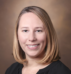
Andreana Holowatyj, PhD, MSCI

“When you take these alarming rates that we are seeing for appendiceal cancer across generations, together with the fact that 1 in every 3 patients diagnosed with appendiceal cancer is diagnosed under the age of 50, these point to a timely need for everyone to be aware of the signs and symptoms of appendix cancer.

“Albeit cancer of the appendix is rare, it is important for individuals who have these symptoms to see a health care professional. Ruling out the possibility of an appendix cancer diagnosis, or diagnosing it early, is important for this cancer as we continue to learn what factors may be contributing to this worrisome trend,” Holowatyj said.

Symptoms may include abdominal pain, bloating, changes in bowel habits, and loss of appetite. 

Appendiceal cancer is a rare cancer with about 3,000 new cases diagnosed every year in the United States, according to prior studies, but Holowatyj and colleagues took a closer look at statistics by combing through the SEER database. 

“As incidence rates in younger generations are often indicative of future disease burden, these results support the need for histology-specific investigations of appendiceal adenocarcinoma, as well as increased education and awareness of appendiceal adenocarcinomas among health care providers and the public,” the study stated. 

Appendiceal cancer has had no standardized screening guidelines, risk factors or tumor classifications — a lack of clinical evidence that has resulted in late diagnosis and poor prognosis. Up to 1 of every 2 patients is diagnosed with distant metastatic disease, and five-year survival rates vary between 10% and 63%.  

Earlier this year, Holowatyj and a team of experts identified six key research priority areas to deliver a fundamental understanding of appendiceal tumors and to improve treatments and outcomes for patients.  

The recommendations published Feb. 20 in Nature Reviews Cancer are the result of a concerted focus by the Appendix Cancer Pseudomyxoma Peritonei (ACPMP) Research Foundation to better understand the disease that afflicts an estimated 3,000 new patients across all age groups each year. 

The recommendations arose from the inaugural ACPMP Research Foundation Scientific Think Tank, sponsored by ACPMP and chaired by Holowatyj at Vanderbilt-Ingram Cancer Center in December 2023. 

The current study in the Annals of Internal Medicine received support from ACPMP and the National Institutes of Health (grants K12HD043483 and P50CA236733). 

Other authors on the study are Mary Kay Washington, MD, PhD, professor of Pathology, Microbiology and Immunology at VUMC, Richard Goldberg, MD, of the West Virginia University School of Medicine, and Caitlin Murphy, PhD, MPH, of the UT Health Houston School of Public Health. 

The post Study shows sharp increase in appendix cancer for Generation X and millennials  appeared first on VUMC News.

AI researchers develop new technologies for cancer care  

Submitted by vicc_news on

Researchers at Vanderbilt University Medical Center using artificial intelligence have helped develop two technologies for improving cancer care. 

One technology called MSI-SEER, described in a study published in npj Digital Medicine, better predicts microsatellite instability-high status from standard pathology slides and provides clinicians with specific data, including any uncertainties with predictions. The other technology, a breakthrough three-dimensional imaging tool described in a study published in Nature Communications, has transformative potential beyond cancer diagnostics. 

These new technologies showcase how VUMC researchers are using the power of AI to meet a wide range of medical needs, said Tae Hyun Hwang, PhD, professor of Surgery, founding director of the Molecular AI Initiative, and director of AI Research for the Vanderbilt Section of Surgical Sciences. He noted that the 3D imaging could significantly advance development of therapeutic drugs, provide more detailed assessments of organ transplant rejections, assist with personalized medicine, and aid with tissue analysis for biopharmaceutical development. 

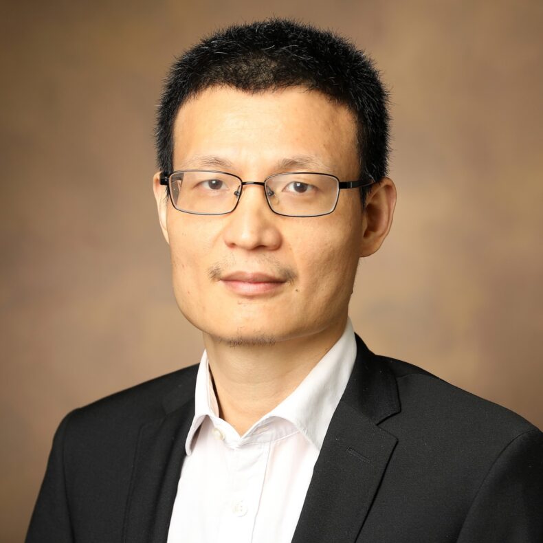
Tae Hyun Hwang, PhD
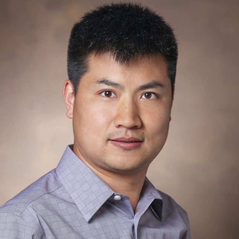
Tae Hyun Hwang, PhD

“This technology fundamentally redefines how we visualize and analyze tissue architecture, moving from traditional two-dimensional views to full 3D microenvironment mapping at the subcellular level,” said Hwang, a corresponding author of the study, who provided senior leadership in the development, validation and translational development of the technology. 

The 3D study published in Nature Communications introduced an innovative framework that integrates holotomography with deep learning to generate hematoxylin- and eosin-stained images directly from thick tissue samples. This noninvasive, AI-driven approach preserves tissue integrity, overcomes the traditional 4- to 5-micron thickness limit of routine histology, and enables volumetric visualization of biological structures up to 50 microns thick. 

By preserving tissue samples and avoiding chemical alteration, this method also ensures compatibility with downstream molecular assays, such as spatial transcriptomics, proteomics and genomic profiling — enhancing the breadth and depth of diagnostic and research capabilities.  

“This is not just a digital copy of hematoxylin- and eosin-staining,” Hwang said. “It is a foundational platform for AI-driven volumetric tissue analysis that accelerates discoveries in oncology, immunology, regenerative medicine and therapeutic development.” 

The multi-institutional effort also included researchers from KAIST, Tomocube Inc., Yonsei University College of Medicine and Mayo Clinic. Hwang received funding support from the National Cancer Institute (grants R01CA276690, R37CA265967, U01CA294518). 

VUMC researchers developed the MSI-SEER predictor technology in collaboration with Mayo Clinic, Yonsei Severance Hospital and Seoul St. Mary’s Hospital in South Korea. This technology identifies patients who will benefit from an immunotherapy that might otherwise be missed with existing prediction models. 

Microsatellite instability-high (MSI-H) status is a well-established biomarker used to identify patients likely to respond to immune checkpoint inhibitors, especially patients with gastrointestinal cancers. However, traditional testing methods — including immunohistochemistry and PCR-based assays — offer only a binary result and often miss focal or heterogeneous MSI-H regions within tumors.  

MSI-SEER overcomes this limitation by dividing each pathology slide into thousands of image tiles and generating region-by-region predictions of MSI-H probability. This enables visualization of the tumor’s spatial heterogeneity and quantification of the MSI-H fraction across the tumor. In multiple cases, MSI-SEER identified MSI-H regions in tumors previously classified as microsatellite stability, and those patients subsequently responded to immunotherapy. 

“This is analogous to what we say in HER2-low gastric cancer, where patients previously not eligible for targeted therapy are now being treated with agents like trastuzumab deruxtecan,” Hwang said. “Likewise, patients with low or heterogeneous MSI-features may now be reconsidered for immunotherapy if spatially resolved analysis like MSI-SEER is used.” 

A key innovation of MSI-SEER is its ability to report not only predictions but the confidence level for each result.  

“AI should not dictate clinical decisions; it should support them,” Hwang said. “MSI-SEER gives clinicians both the answer and a measure of how reliable the answer is. It’s not about replacing human expertise but about combining the best of AI computation with physician judgment to drive safe, precise decisions.” 

Hwang, who conceptualized the study and is the paper’s senior author, received research support from the National Cancer Institute and the Department of Defense. He also received support from the Eric and Wendy Schmidt Fund for AI Research and Innovation and the American Association for Cancer Research Innovation and Discovery Grant.  

Other VUMC researchers who authored the study are Sunho Park, PhD, Minji Kim, MS, Jean Clemenceau, PhD, and Inyeop Jang, PhD. 

The post AI researchers develop new technologies for cancer care   appeared first on VUMC News.

Andreana Holowatyj named ‘40 Under 40 In Cancer’ winner

Submitted by vicc_news on
Andreana Holowatyj, PhD, MSCI

Andreana Holowatyj, PhD, MSCI, assistant professor of Medicine, has been named to the 40 Under 40 In Cancer Class of 2025 by the Association for Value-Based Cancer Care.

This year’s class was selected from more than 3,000 nominations. The awards initiative identifies and recognizes contributions across the field of cancer by rising stars and emerging leaders under the age of 40. 

Holowatyj’s research is focused on early-onset cancers, including colorectal and appendiceal cancers. She has received the National Cancer Institute’s Method to Extend Research in Time (MERIT) Award to support her ongoing investigation into how early-onset colorectal cancer and its treatments impact reproductive health. MERIT Awards provide longer-term funding than is typical for most grants to early-stage investigators whose research competence and productivity are distinctly superior and who are highly likely to continue to perform in an outstanding manner. As part of this Award, Holowatyj established and leads the Preserving Fertility After Colorectal Cancer (PREFACE) clinical study, which is currently recruiting patients.  

She is focused on providing evidence-based guidance that will ultimately improve clinical care and outcomes for individuals ages 18 to 49 when diagnosed with cancer. Holowatyj and her team discovered that 1 in 2 young cancer patients report that a health care provider involved in their cancer care did not discuss options to preserve fertility prior to starting cancer treatment. 

Her research has been published in high-impact medical research journals and has led to clinical practice changes and revisions to consensus guidelines. She has also been invited to serve on several international committees, including the American Joint Committee on Cancer Lower Gastrointestinal Tract Expert Panel that updates clinical cancer staging systems, the Fight Colorectal Cancer Global Early-Onset Colorectal Cancer Think Tank, and as the inaugural chair of the Scientific Advisory Board for the Appendix Cancer Pseudomyxoma Peritonei (ACPMP) Research Foundation. This year, with the support of the ACPMP Research Foundation, she led an expert recommendation report in the journal Nature Reviews Cancer that identified six key research priority areas to deliver a fundamental understanding of appendiceal tumors and to improve treatments and outcomes for patients with this rare cancer.

The post Andreana Holowatyj named ‘40 Under 40 In Cancer’ winner appeared first on VUMC News.

Study finds navigational bronchoscopy as effective and safer alternative to transthoracic biopsy for lung nodules  

Submitted by vicc_news on

Navigational bronchoscopy is as effective as the traditionally used transthoracic needle biopsy for diagnosing lung nodules, but with significantly fewer complications, per a new study published May 18 in the New England Journal of Medicine. This development may shift medical practice and reduce hospitalizations for patients undergoing lung nodule biopsies, said researchers.  

Each year, millions of lung nodules are detected during routine X-rays or CT scans. While most nodules are benign, some are cancerous and when found at this stage they are the earliest and most curable stage of lung cancer.   

Accurate biopsy of lung nodules is often required to tell benign nodules from malignant ones. To date, the most commonly used method, CT-guided transthoracic needle biopsy, carries a high complication rate — including pneumothorax, or partial lung collapse, affecting about 25% of patients. Treatment of the partially collapsed lung often requires a chest tube during a multiple day inpatient stay.  

Robert Lentz, MD
Robert Lentz, MD

This study is the first to directly compare these biopsy techniques and the results are compelling, said first author Robert Lentz, MD, associate professor of Medicine and Thoracic Surgery in the Division of Allergy, Pulmonary and Critical Care Medicine at Vanderbilt University Medical Center. The study, conducted across seven centers in the United States, compared the two techniques in a multicenter, randomized trial involving 234 patients with lung nodules between 10-30 mm.  

Navigational bronchoscopy uses a sophisticated targeting system and 3D imaging to guide biopsy tools through small peripheral airways directly to a lung nodule.   

The comparison revealed a diagnostic accuracy of 79% using navigational technology, closely matching the 74% accuracy of transthoracic biopsy. More importantly, the risk of pneumothorax was significantly lower, occurring in only 3% of bronchoscopy patients compared to 35% in those undergoing transthoracic biopsy. Severe cases requiring hospital admission or chest tube insertion were less than 1% with bronchoscopy, as opposed to 14% with the traditional method.  

“With approximately 300,000 lung nodule biopsies performed annually in the U.S., shifting to navigational bronchoscopy could greatly reduce complications and hospital stays. This study confirms its diagnostic efficacy and superior safety profile, making it the preferred choice for lung nodule diagnosis,” said senior author Fabien Maldonado, MD, MSc, professor of Medicine and Thoracic Surgery and director of Interventional Pulmonology.  

The research team plans to continue exploring optimal biopsy techniques, comparing different navigational and robotic bronchoscopy systems, and studying novel biopsy tools, said Maldonado, Pierre Massion Director in Lung Cancer Research.  

They are also involved in research on bronchoscopic ablation of lung cancer, advancing the field of interventional pulmonology.  

“As a relatively new medical subspecialty, we are quite proud and excited to have matured our research infrastructure to the point of being able to produce high quality multicenter trials like this, and hope that publication of this trial will serve as an introduction of sorts for interventional pulmonology to the larger general medical audience,” said Lentz.   

The post Study finds navigational bronchoscopy as effective and safer alternative to transthoracic biopsy for lung nodules   appeared first on VUMC News.

VoLo Foundation grant supports Southern Environmental Health Study

Submitted by vicc_news on

A grant from VoLo Foundation will augment federal funding for the Southern Environmental Health Study, an initiative to determine whether environmental exposures are contributing to cancer cases in the region.

The study received its initial funding from the National Cancer Institute and the Vanderbilt-Ingram Cancer Center. A grant from VoLo Foundation — a private nonprofit organization dedicated to accelerating global impact through science-based solutions, education enhancement, and health improvement initiatives — will allow investigators to recruit additional participants and conduct methylation- and proteomics-based biological aging assays to check for biomarkers of early disease risk.

On cancer incidence and mortality maps, some of the highest rates in the country appear in the southern U.S., but whether environmental exposures are a contributing factor has not been scientifically determined. The study is a long-term cohort investigation that will follow participants for at least 10 to 20 years. The participants are between the ages of 40 and 70 and live in Alabama, Arkansas, Delaware, Florida, Georgia, Kentucky, Louisiana, Maryland, Mississippi, Missouri, North Carolina, Oklahoma, South Carolina, Texas, Tennessee, Virginia, West Virginia or the District of Columbia.

“Humans are exposed to large numbers of chemicals and their mixtures with more than 80,000 chemicals having been registered by the Environmental Protection Agency,” said Wei Zheng, MD, PhD, MPH, the Anne Potter Wilson Professor of Medicine and the director of the Vanderbilt Epidemiology Center. “However, very few of them have been adequately investigated in relation to human cancers and other diseases in epidemiologic studies. There are considerable challenges in studying environmental exposures in epidemiologic studies.”

Participants will wear silicone wristbands designed to collect chemicals, which researchers will use to measure exposure to approximately 500 compounds. They will also analyze blood samples to assess the internal exposome using both liquid chromatography and gas chromatography with high-resolution mass spectrometry, allowing them to detect approximately 1,500 confirmed chemicals and endogenous metabolites. To identify potential environmental carcinogens, the researchers will use an innovative exposome-wide association study to link chemical exposures with disease biomarkers, including indicators of biological aging and inflammation.

“Supporting the Southern Environmental Health Study aligns with our mission to advance data-driven solutions that can lead to healthier, more resilient communities. This research has the potential to uncover possible links between environmental exposures and chronic diseases,” said David Vogel, co-founder and chief scientist of VoLo Foundation.

Vogel and his wife, Thais Lopez Vogel, formed the VoLo Foundation in 2014.

“We are very grateful for the support of the VoLo Foundation and believe that this study will generate significant amounts of novel data regarding potential impacts of environmental exposures on human health and pave the way for future studies in this important area,” Zheng said.

The post VoLo Foundation grant supports Southern Environmental Health Study appeared first on VUMC News.

All cancer patients at Vanderbilt-Ingram will have the opportunity to ring the bell 

Submitted by vicc_news on

From his infusion chair, Matt Duckworth would clap and yell “woohoo” each time he heard another cancer patient ring the bell to mark the completion of chemotherapy treatment, even though he knew he would never get that opportunity. 

Matt Duckworth
Matt Duckworth

On Friday, April 25 — seven months after his death — the bell pealed loudly at the Vanderbilt-Ingram Cancer Center infusion clinic as a plaque was unveiled in his honor and the announcement was made that patients receiving palliative chemotherapy would also get to ring the bell going forward. 

Duckworth was in the prime of his life at age 41, working as director of Population Health Operations for Vanderbilt Health Affiliated Network and doting on his three dogs with his wife, Suzanne, when he was diagnosed in 2020 with Stage 4 gastroesophageal cancer that had metastasized to his liver.  Although the chemotherapy didn’t cure his cancer, the treatments did extend his life for four years. 

“Matt had infusions weekly, and as we were sitting in the infusion room for hours at a time, we would hear the bell ring, and we would hear everybody clapping,” said Suzanne Duckworth. “He would clap and yell, ‘Woohoo!’ He just emitted such a positive vibe and aura. I would sit there and think, ‘He’s never going to get that because he has terminal cancer.’ Now, that thought might have come into his brain, but you would never know it because he never said it.” 

Suzanne Duckworth consoles her mother-in-law, Betsye Duckworth, as Stephanie Broderick, who worked with Matt Duckworth, reads words of appreciation from his colleagues. (photo by Susan Urmy)

She asked Vanderbilt-Ingram about adding a plaque, and a ceremony was held in Matt’s memory to mark the occasion. Colleagues, family and friends shared stories about the man, who was beloved for his sense of humor, love for trips to Disney World and his affinity for dogs. His physician, Michael Gibson, MD, PhD, associate professor of Medicine, spoke about the relationship he developed with Matt and how much he enjoyed spending time with him.  

Suzanne Duckworth stated, “This is for you, Matt,” and rang the bell. 

His mother, Betsye, stood nearby wiping away a tear. On the plaque that had just been unveiled was a poem written by his sister, Jill Comfort.

I stand before the bell today 

Not for an end, but to proudly say 

I fought with every breath I had  

Through days of pain and times of glad 

I ring this bell for all I’ve done 

For every battle, fought and won 

For every day, for every try 

I ring the bell — head held high 

Ten years younger than Matt, Comfort had come to Nashville from Mississippi, to help Suzanne, who is a Vanderbilt nurse, with her big brother when he began hospice care. It had also bothered her that Matt never got to ring that bell, so she bought him one and read him the poem she had written.  

“But he wouldn’t ring the bell because that was admitting defeat,” Suzanne Duckworth said. 

A native of Clinton, Mississippi, Matt excelled in life, attaining an undergraduate degree in journalism, a Master of Science in public relations and then a Juris Doctor from Mississippi College of Law with honors followed by a Doctor of Healthcare Administration.  

He and Suzanne married in 2003 and eventually moved to Nashville, where they both established careers at Vanderbilt University Medical Center.  

“When things were running down, he didn’t want to give up,” Comfort said. “Then one day, he asked me if I would read the poem at his funeral and ring the bell. That somehow transpired into where we are today. We talked about how there were other people in his situation that don’t get that opportunity who are needing treatments for years and years. They need some encouragement.” 

Suzanne Duckworth approached Julie Bulger, manager of Patient- and Family-Centered Care at Vanderbilt-Ingram about allowing any chemotherapy patient to ring the bell regardless of whether they had completed treatment or achieved remission. 

“When Suzanne shared her idea about a new plaque, I deeply appreciated what she said, how when Matt heard the bell, he felt happy for those who rang it,” Bulger said. “It is rung traditionally at the end of treatment, but many patients may never have that opportunity. This thought comes from a lot of places, not only from Matt, but Suzanne articulated it so beautifully.  I believe this is going to resonate with many people.

“The bell can be rung at any time. It doesn’t have to be the last day of treatment. It can be rung because you’re having a bad day, and you’re fighting, and it’s hard. Or you have achieved a different milestone. You want to ring that bell because it’s going to mean something for you, and you want to feel uplifted,” Bulger said.

The post All cancer patients at Vanderbilt-Ingram will have the opportunity to ring the bell  appeared first on VUMC News.

Vanderbilt-Ingram 26th Annual Scientific Symposium focuses on AI 

Submitted by vicc_news on

Experts on the research, clinical use, governance and ethical use of artificial intelligence gathered for the recent Vanderbilt-Ingram Cancer Center 26th Annual Scientific Symposium. 

In a twist from years past, graduate students and postdoctoral fellows took the helm in selecting the topics and inviting speakers focused on “Artificial Intelligence in Cancer Research and Clinical Care.”

The keynote speakers were Eytan Ruppin, MD, PhD, chief of the Cancer Data Science Laboratory at the National Cancer Institute, and Gelareh Zadeh, MD, PhD, chair of the Department of Neurologic Surgery at Mayo Clinic.  

Ruppin detailed how he is developing computational approaches for advancing precision oncology, and Zadeh explained how she is using integrated multi-platform molecular analysis of brain tumors to predict patients’ responses to targeted therapies. Ruppin participated in panel discussions about artificial intelligence. 

“I am enriched talking to you guys,” Ruppin said. “I develop AI materials, but I am not using them to treat patients. I am learning a lot.” 

Douglas Flora, MD, executive medical director of Oncology Services at the Yung Family Cancer Center at St. Elizabeth in Edgewood, Kentucky, and the editor-in-chief of AI in Precision Oncology, replied, “All of us are cross pollinating. That’s why I love a symposium like this.” 

In opening the first panel discussion that focused on ethics, Ellen Wright Clayton, MD, JD, the Craig-Weaver Professor of Pediatrics, professor of Law and professor of Health Policy at Vanderbilt, framed artificial intelligence from an historical perspective, noting that “decision support is not new to medicine.” She gave specific examples of how clinicians can use artificial intelligence for decision support but stressed that they should not rely solely on it for treatment plans. 

“It is not OK simply to get the AI output and just do what it says,” Clayton said. “Maybe it is OK, but it is always required to see if that’s the right advice. Always.” 

In another twist from years past, the Mission Moment, which is a personal testament from a patient, was presented by a pediatric cancer survivor for the first time. Easton Reeder, 13, who has undergone surgery and chemotherapy for pilocytic astrocytoma — a type of brain tumor — shared about his experiences living with cancer. He told his story vividly with flashes of humor, describing how being tossed in the air like a rodeo clown by a Great Dane led to his diagnosis.

Clinicians initially concluded that he had a concussion because of persistent headaches that followed, but his mother, who is a nurse, insisted on a brain scan. Reeder, a committed athlete, who continued playing sports even while undergoing chemotherapy, was given a jersey signed by Vanderbilt baseball players.

“I learned that tomorrow isn’t a promise, and I have to make the best of every moment I have,” Reeder said. “I also learned that there is no ‘normal button.’ I have been trying to learn that power since forever, until I realized that power is not to be . . . God has proven to me that anything is possible through him.” 

In his welcoming remarks, Vanderbilt-Ingram director Ben Ho Park, MD, PhD, emphasized the importance of training new generations of cancer researchers and clinicians. 

“This is an opportunity for us to celebrate all the cancer research going on at Vanderbilt-Ingram,” Park said. “For all of you who don’t know, we really run the spectrum of everything research: clinical, population science, laboratory science and everything in between. This is our time of the year when we get to showcase and highlight not only the great science that our external panelists and presenters are going to bring — but you will be duly impressed, as I always am, by what our trainees bring to the table. The future really is bright, and we have to keep sustaining our future by encouraging and mentoring the next generation, which will ultimately lead to more cures.” 

From left are Christopher Williams, MD, PhD, associate director of Research Education at VICC, Michael Robinson, MD; Guochong “Damon” Jia, PhD, postdoctoral scholar of the year, Katie Brown, PhD, co-chair of the Vanderbilt-Ingram Scientific Symposium, Candace Grisham, MS, co-graduate student of the year, Xiaopeng Sun, PhD, co-graduate student of the year, Jared Rhodes, co-chair of the Scientific Symposium, Ben Ho Park, MD, PhD, director of VICC. (photo by Donn Jones)

Two tied in voting for the Graduate Student of the Year. Candace Grisham, MS, received the honor for her research into brain tumors, including a study she authored that was published in Clinical Neurology and Neurosurgery. Xiaopeng Sun, PhD, is the other co-awardee for his research into biomarkers to predict immunotherapy outcomes in patients, and his prolific contributions to that field of study, including 12 studies published in scientific journals. 

Guochong “Damon” Jia, PhD, MPH, is the Postdoctoral Scholar of the Year. He was selected for his high-impact research that has advanced the understanding of cancer genetics and epidemiology, including the largest genetic study ever conducted on breast cancer in African ancestry populations, which was published in Nature Genetics

The poster exhibition was one of the largest ever for the annual event. Sarah Reed took home the overall winner award for her entry “Identifying Genotype-Specific Effects of CHIP on Solid Tumors Using Chimeric Mouse Modeling and Clinical Data.” 

In the Translational Science Category, Julia Steele won first place; Alexander Kwiatkowski, PhD, won second place; Heather Beasley, PhD, won third place; and honorable mention went to Jacey Marshall. 

Robust participation in the Basic Science Category resulted in duplicate prizes due to the number of entries. Rachel Sinard and Lincoln Brown won first place prizes. Emily Green and Logan Vlach received second place awards, andAnna Gilbert and Alyssa Jarabek received third place awards. Honorable mentions went to Sydney Bates, Nicholas Eleuteri, Sarah Glass, PhD, Gabriela Gonzalez Vasquez, Maxwell Hamilton, Yash Pershad, Jared Rhodes and Brenda Rios. 

In the Population Science Category, Michael Betti received first place; Duc Huy Le, MD, MBA, received second place; and there was a tie for third place with Melissa Goldin and Jiajun Shi, PhD, both receiving awards. Honorable mention went to Grace Xu. 

The post Vanderbilt-Ingram 26th Annual Scientific Symposium focuses on AI  appeared first on VUMC News.

Three Vanderbilt-affiliated cellular therapy programs receive reaccreditation

Submitted by vicc_news on

The cellular therapy programs at Vanderbilt-Ingram Cancer Center, Monroe Carell Jr. Children’s Hospital at Vanderbilt, and the VA Tennessee Valley Healthcare System have received reaccreditation from the Foundation for the Accreditation of Cellular Therapy (FACT).

Founded in 1995, FACT establishes standards for high quality medical and laboratory practice in cellular therapies. FACT is a nonprofit corporation co-founded by the International Society for Cell and Gene Therapy and the American Society for Transplantation and Cellular Therapy for the purposes of voluntary inspection and accreditation in the field of cellular therapy.

“FACT is an internationally recognized accrediting body for hospitals that offer stem cell transplant and cellular therapy, and recognition by FACT indicates that the accredited institution has met the most rigorous standard in every aspect of cellular therapy,” said Adetola Kassim, MD, professor of Medicine and director of the Vanderbilt Stem Cell Transplant and Cellular Therapy Program. “This covers the entire spectrum of stem cell therapy from clinical care to donor management, cell collection, processing, storage, transplant, administration and cell release.”

The Vanderbilt and VA Tennessee Valley Healthcare System programs received accreditation notification on April 14 after on-site inspections in October 2024. The accreditation is effective for three years.

“Congratulations to the entire Vanderbilt University Medical Center and VA Tennessee Valley Healthcare System team on achieving FACT accreditation and providing quality care for the people we serve,” Kassim said.

The post Three Vanderbilt-affiliated cellular therapy programs receive reaccreditation appeared first on VUMC News.

Subscribe to Cancer