This is a first-in-human, multicenter, open-label, phase 1 study to evaluate the safety, PK, PD and preliminary efficacy of STX-0712 in patients with advanced CMML and AML for whom there are no further treatment options known to confer clinical benefit.

Study led by Monroe Carell Jr. Children’s Hospital at Vanderbilt looks at international cohort with rare cancer

Submitted by vicc_news on
This is a de-identified MRI scan from the REDCap database depicting numerous discrete lesions including fourth ventricle, thalamus, midbrain, cerebellum, brainstem and spine, highlighting the disseminated disease. (image courtesy/Michael Dewan, MD)

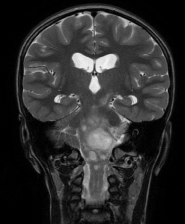
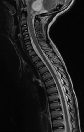
Pediatric-type, low-grade gliomas (PLGG) are the most common central nervous system (CNS) tumor in children, but in rare cases the tumors spread — known as disseminated pediatric low-grade gliomas (DPLGGs) — which leads to increased morbidity and mortality.

Researchers, led by Michael Dewan, MD, MSCI, compiled an international cohort of more than 260 DPLGG patients from 39 sites and 13 countries to study why low-grade brain tumors behave aggressively, as well as to identify the causative genetic alteration. Study findings were recently published in Neuro-Oncology.

“Our findings will help predict which children are at an elevated risk of developing disseminated low-grade gliomas as well as serve as a guide toward a more effective, personalized treatment,” said Dewan, associate professor of Neurological Surgery and Pediatrics at Monroe Carell Jr. Children’s Hospital at Vanderbilt and one of the study’s senior authors.

This is a de-identified MRI scan from the REDCap database depicting numerous discrete lesions including fourth ventricle, thalamus, midbrain, cerebellum, brainstem and spine, highlighting the disseminated disease. (image courtesy/Michael Dewan, MD)

“Not all childhood brain tumors that look low-grade act the same,” he said. “This study expands our understanding of the clinical, pathologic and molecular features of this challenging disease. We found that when these tumors spread early or widely — especially in young children — outcomes are worse, but newer, targeted drugs work better than standard chemotherapy for many patients.”

Study findings inform prognosis and treatment decisions, particularly by supporting the earlier use of targeted, less-invasive therapies that may enhance survival and reduce treatment toxicity for children.

“This research improves our clinical and biological understanding of this rare disease, provides insights for improving patient care, and directs future clinical trials and basic science research,” said Dewan.

The post Study led by Monroe Carell Jr. Children’s Hospital at Vanderbilt looks at international cohort with rare cancer appeared first on Vanderbilt Health News.

Vanderbilt Health and Bertis establish collaboration for cancer drug discovery

Submitted by vicc_news on

Vanderbilt Health and Bertis, an artificial intelligence-driven proteomics-based precision medicine company, have announced a joint research and co-development collaboration. The endeavor marks a significant milestone in oncology by advancing the convergence of AI, spatial biology and translational cancer research.

By integrating Vanderbilt Health’s Molecular AI Initiative capabilities with Bertis’ proprietary deep proteomics and AI-enabled target discovery technologies, the collaboration will build an advanced, spatially resolved dataset to identify novel therapeutic targets and predictive biomarkers.

Traditional target discovery often relies on bulk tissue analysis, which loses the critical context of how cells are organized within a tumor. Vanderbilt Health’s Molecular AI approach changes this paradigm by employing sophisticated computational spatial analysis to generate high-resolution spatial molecular maps. This AI-driven spatial biology allows researchers to visualize and decode the complex architecture of the tumor microenvironment, specifically identifying how tumor, immune and stromal (connective tissue) cells interact in biologically and therapeutically relevant regions. By mapping the precise locations and spatial relationships of these cells, the Molecular AI platform can isolate the key cell populations responsible for treatment response or resistance.

These advanced spatial insights are then integrated with Bertis’ cutting-edge proteomics capabilities. While Vanderbilt Health maps the critical spatial context, Bertis will conduct deep proteomic and metabolomic profiling, applying its proprietary AI-enabled computational models to prioritize the most viable, druggable targets.

Tae Hyun Hwang, PhD
Tae Hyun Hwang, PhD

The initial focus of this joint research will be on HER2-low tumors (cancers that express low levels of the growth-promoting protein HER2), a historically challenging clinical area, with the potential to expand into additional tumor types based on data outcomes and joint scientific discussions. By layering spatial context over proteome-level data, the teams aim to pinpoint cell surface proteins that are uniquely positioned for emerging therapeutic modalities, including antibody-drug conjugates and cell-based therapies.

This sophisticated AI-driven spatial multimodal and deep proteomics pipeline is spearheaded by Tae Hyun Hwang, PhD, professor of Surgery, founding director of Molecular AI Initiative and director of AI Research in the Section of Surgical Sciences at Vanderbilt Health. Hwang also co-leads gastric cancer atlas efforts within the National Cancer Institute-funded Human Tumor Atlas Network (HTAN) and is spearheading international HTAN collaborations with South Korea’s National Cancer Center.

Highlighting the clinical necessity of this integrated approach, Hwang said, “Identifying therapeutic targets and understanding treatment response require a precise view of proteins, spatial context and tumor biology. By combining Vanderbilt Health’s Molecular AI and spatial analysis capabilities with Bertis’ proteomics and AI-enabled target discovery platform, this collaboration is designed to generate high-confidence therapeutic targets and predictive biomarkers that can support future translational research and therapeutic development.”

Bertis is led by co-CEOs Dong-young Noh and Seung-man Han, who emphasized the collaboration accelerates the global reach of their platform.

“Collaborating with Vanderbilt Health, a leading U.S. academic medical center with strong expertise in Molecular AI, spatial biology and cancer research, is highly meaningful and reflects the growing global recognition of Bertis’ technological capabilities,” Han said. “Through this collaboration, we aim to expand the role of AI-driven proteomics in drug discovery and identify therapeutic targets that may open new possibilities in oncology.”

The post Vanderbilt Health and Bertis establish collaboration for cancer drug discovery appeared first on Vanderbilt Health News.

This study is being done to answer the following questions: Can we lower the chance of your gastric cancer from growing or spreading by administering paclitaxel chemotherapy directly into your abdominal cavity in addition to chemotherapy given through a vein in your arm? Will administering paclitaxel chemotherapy directly into your abdominal cavity, in addition to chemotherapy given through a vein in your arm help you live longer? We are doing this study because we want to find out if this approach is better or worse than the usual approach for your gastric cancer. The usual approach is defined as care most people get for gastric cancer. If you decide to take part in this study, you will first receive a surgical procedure called a diagnostic laparoscopy. This will help the study doctors learn more about your gastric cancer. Laparoscopy is a minimally invasive surgery for which you will be placed under general anesthesia. Then the surgeon will make small incisions (5mm) on your belly through which a camera and thin instruments are introduced to evaluate the abdomen. This procedure takes about 1 hour to complete. Your study group will be assigned during the surgery. The study groups are described further in the 'What are the study groups?' section below. If you are placed into the study group 1, you will not have an intraperitoneal port (a small device which is placed under the skin and fat of your upper abdomen and a tube that is placed into the abdomen). If you are placed into the study group 2, you will have an intraperitoneal port placed. The reason is that in addition to standard chemotherapy, which is given through a vein in your arm, this port will be used to deliver the medication paclitaxel directly inside your abdomen when you are ready to start study treatment. It is important to know that you will not know your study group until after the surgery is over. This is because information that is learned during the surgery will help determine which study group you are put in. Once you have fully healed from this surgery, you will start study treatment. Depending on which study group you are assigned, you will either receive a standard chemotherapy regimen (the regimen will be chosen by you and your doctor) if you are in study group 1, or paclitaxel through a tube in your belly plus chemotherapy given through a vein in your arm if you are in study group 2. All participants will get treatment for three (3) months after which you will undergo reevaluation. If the disease is under control or responding to treatment, you may continue the assigned treatment until your disease gets worse, the side effects become too severe, or you may be offered a surgical procedure to remove the cancer if the amount of disease is low and can be completely removed as determined by a surgeon. There is a very small chance that during the laparoscopy surgical procedure, the doctor might find something called "intra-abdominal adhesions". These are areas where the stomach has healed previously and created scar tissue. If this scar tissue prevents the surgeon from being able to place a port in the correct area, you would be ineligible to receive the study treatment. If this happens, you may still receive standard of care therapy after your surgery, but you will not be able to continue on the study. If you have more questions about this, you can ask your surgeon or the study team to help. After you finish your study treatment, your doctor or study team will watch you for side effects. They will continue to follow your condition every three (3) months during the first two (2) years, then every six (6) months until year 5. You may be reevaluated with Chest/Abdomen/Pelvis scans every three-six (3-6) months for up to five (5) years if decided by your doctor.
This three-arm randomized phase 2 trial will enroll advanced clear cell RCC patients (all IMDC risk groups). Patients will be randomized 2:2:1 to either Arm A (fianlimab/ cemiplimab/ ipilimumab), Arm B (fianlimab/ cemiplimab), or Arm C (standard ipilimumab/ nivolumab), respectively.

Demographic and histologic factors influence risk of colon polyp recurrence

Submitted by vicc_news on

When colorectal adenomas — polyps that can develop into colon cancer — are removed during a colonoscopy, patients look for guidance about when to have another colonoscopy to look for new growths.

Current surveillance guidelines emphasize polyp characteristics, but a new study led by Vanderbilt Health researchers demonstrates that demographic factors also influence colorectal adenoma recurrence. Their findings, reported in the journal JAMA Network Open, underscore the need for surveillance strategies that factor in polyp characteristics and population-specific risk profiles, as well as variation over time.

Colorectal cancer is the second most common cause of cancer-related death in the United States, according to the National Cancer Institute. Removing precancerous polyps during colonoscopy procedures significantly reduces the burden of colorectal cancer. But about 30% of patients who have a colorectal adenoma removed will develop recurrent adenomas, putting them at increased risk for developing cancer.

Xingyi Guo, PhD

“The current guidelines for surveillance after polyp removal stratify recurrence risk primarily based on polyp characteristics including size, histology and number,” said Xingyi Guo, PhD, associate professor of Medicine in the Division of Epidemiology and co-corresponding author of the new study. “The guidelines universally neglect demographic variables such as race, sex, family history, age and obesity, and may therefore be inadequately addressing population heterogeneity in recurrence risk and timing.”

The researchers, led by Guo and co-corresponding author Zhijun Yin, PhD, associate professor of Biomedical Informatics, conducted a retrospective cohort study using Vanderbilt Health electronic health records for about 3.5 million patients.

Zhijun Yin, PhD

They identified a study group of 59,667 adult patients who had an initial colonoscopy with polyp removal between January 1990 and July 2024, and up to 25 years of follow-up. About 29.5% of the patients experienced adenoma recurrence within five years.

The researchers evaluated associations between recurrence and demographic variables: race and ethnicity, sex, obesity (body mass index greater than 30), family history of colorectal cancer or polyps, and age at time of initial adenoma (younger than 50 versus 50 and older). They examined associations with adenoma features including histology, size, number and dysplasia (abnormal cell structure).

Among their notable findings:

  • High-grade dysplasia had the largest association with early (less than five years) adenoma recurrence, but not with mid- (five to 10 years) or late-term (10 or more years) recurrence.
  • Villous histology exhibited a biphasic pattern: an early elevation and late-term resurgence in recurrence risk.
  • Obesity conferred persistent risk across all surveillance intervals.
  • Female patients with high-risk adenomas had marked late-term risk, exceeding male patients.
  • Colorectal adenoma recurrence demonstrated distinct heterogeneity over time, rather than constant risk.

The researchers noted that the study cohort’s predominantly non-Hispanic white population (87.2%) may limit generalizability of the findings, and other factors that could affect risk like socioeconomic status and lifestyle exposures (e.g., tobacco and alcohol use, physical activity and diet) were not consistently available in health records and could not be included in the analysis.

“Our findings suggest that both histopathologic and demographic factors show time-dependent associations with adenoma recurrence, supporting a shift toward dynamic and individualized surveillance strategies,” Yin said.

Usman Ayub Awan, PhD, epidemiology postdoctoral fellow, and Qingyuan Song, PhD, computer science and biomedical informatics graduate student, are co-first authors of the JAMA Network Open study. Guo, Yin and Wanqing Wen, MD, MPH, research professor of Medicine in the Division of Epidemiology, are co-senior authors of the study. The research was supported by the National Cancer Institute (grants R01CA297582 and R01CA269589).

The post Demographic and histologic factors influence risk of colon polyp recurrence appeared first on Vanderbilt Health News.

RBS2418 is a targeted immune modulator that inhibits ectonucleotide pyrophosphatase/phosphodiesterase 1 (ENPP1). It is designed to promote anti-tumor immunity by preserving endogenous 2'-3' cyclic guanosine monophosphate-adenosine monophosphate (cGAMP) from hydrolysis, thereby activating antigen-presenting cells and promoting robust T cell activation. Ideally, RBS2418 acts synergistically with CTLA-4 inhibitors, such as those in the STRIDE regimen (Tremelimumab plus Durvalumab). The hypothesis is that RBS2418 combined with STRIDE will be safe, well-tolerated, highly immunogenic, and enhance anti-tumor responses in adult participants with advanced, unresectable hepatocellular carcinoma (HCC) compared to STRIDE alone.

Cancer and Environment at Every Scale

Chemotherapy drugs, used in the treatment of cancer, have the potential of inducing peripheral neuropathy (PN) as a side effect. This side effect is commonly referred to as CIPN, or chemotherapy-induced peripheral neuropathy. The Lilac Glove and Boot devices apply a low pressure across the surface of the hands and feet, respectively, to reduce access of chemotherapy to the peripheral nerves on the hands and feet. The small amount of pressure reduces the level of chemotherapy reaching the peripheral nerves, hence increasing the likelihood of nerve preservation during treatment and thus may potentially temporarily prevent the onset of moderate to severe PN symptoms induced by chemotherapy in the hands and feet while receiving treatment
Subscribe to